Boned Human Skull petrous part (of temporal bone)
The ossupra petrosum (O.S.P.) isde-scribed asasmall ossicle, pea-sized atthe most, located on the anterosuperior sur-face ofthe petrous bone, near the tip of this bone andjust anterior and medial to the ganglion ofGasser. Itlies under the dura either independent from itor ad-herent toit. It isusually bilateral and symmetrical and has apparently.
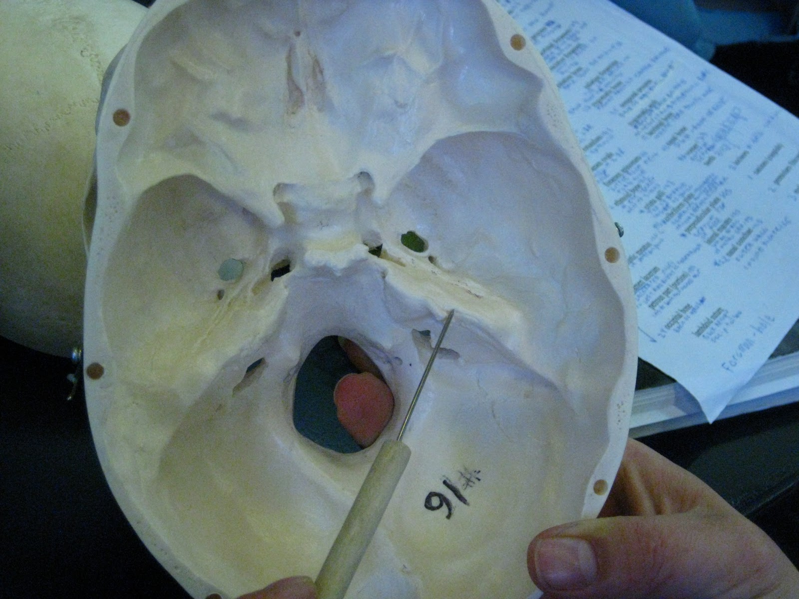
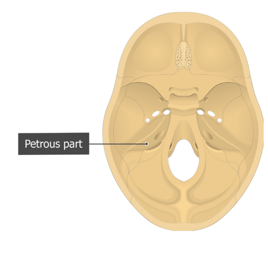
The surface landmarks on the inferior surface of the petrous portion
Inner and middle ear abnormalities (os petrosum) have been studied extensively in humans and mice because of their effect on hearing. Therefore, most anatomic anomalies seen in Chd7‐deficient mice had already been extensively documented in individuals with CHARGE syndrome.
Obecn nauka o kostech Osteologie Rozdlen kost Dlouh
Publicationdate 2016-01-15. This is an updated version of the 2007 article. In this review we present the normal axial and coronal anatomy of the temporal bone by scrolling through the images. Some structures are discussed in more detail with emphasis on related pathology. You will find more temporal bone pathology here.

耳的应用解剖学之颞骨的解剖结构 知乎
Osteolytic lesions on the os petrosum of a Bronze Age individual from La Llana cave (Northern Spain) compatible with a possible case of otitis media. A multifaceted methodological approach. Endocranial view of the individual from La Llana showing the osteolytic lesions on left os temporale. Download : Download high-res image (198KB) Download
PPT Obecná nauka o kostech PowerPoint Presentation ID5090588
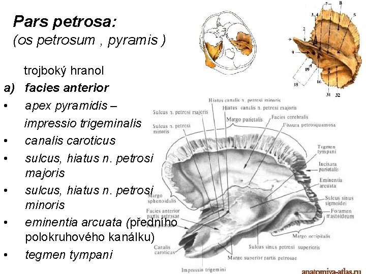
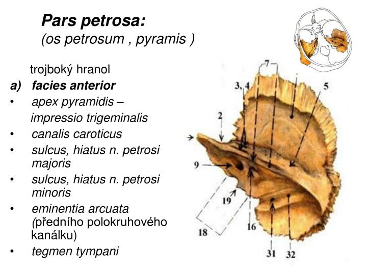
The rock bone (pyramid) is sometimes referred to separately as the bone of the os petrosum. It is a formation similar to a quadrilateral pyramid that protrudes laterally from the back in the ventromedial direction. It contains a complicated cavity space, the so-called labyrinthus osseus (bony labyrinth), in which the sensory organs of hearing.

From Wikiwand Petrous part of the temporal Bones, Sphenoid bone
Pneumatization of the petrous temporal bone apices is an anatomical variant that may be bilateral or asymmetrical. It is important not to confuse it with a pathological lesion especially on MRI. Benign lesions such as cholesterol granuloma and cholesteatoma are more likely to occur in a pneumatized petrous apex. Also, it can be a site for CSF.

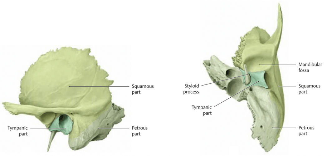
The petrous part of the temporal bone (or more simply petrous temporal
A 53-year-old male patient, with the diagnosis of CG since 2003 with slow progression, presented with diplopia and headache for a couple of weeks. MRI showed a 2.2 × 1.4 cm lesion in the transversal plane above the apex of the right os petrosum. The lesion appeared with a high intensity signal on both T 1 and T 2-weighted imaging.

Os petrosum (equus) Auris media YouTube
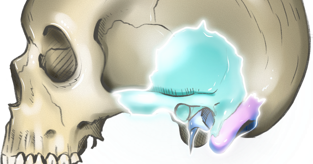
The petrous part of the temporal bone is pyramid-shaped and is wedged in at the base of the skull between the sphenoid and occipital bones.Directed medially, forward, and a little upward, it presents a base, an apex, three surfaces, and three angles, and houses in its interior, the components of the inner ear.The petrous portion is among the most basal elements of the skull and forms part of.

Os petrosum (equus) Canalis facialis YouTube
Temporal bone fracture is usually a sequela of significant blunt head injury. In addition to potential damage to hearing and the facial nerve, associated intracranial injuries, such as extra-axial hemorrhage, diffuse axonal injury and cerebral contusions are common. Early identification of temporal bone trauma is essential to managing the.
Os temporale Axon
Abstract. The anatomy of the petrous apex is described, a system for classifying petrous apex lesions is presented, and commonly encountered petrous apex lesions are discussed, with emphasis on clinical features, CT and MR imaging findings, and normal anatomic variants that may mimic disease. The petrous apex is a complex region of the central.
CTos petrosum
Het rotsbeen, steenbeen, os petrosum of pars petrosa ossis temporalis is een onderdeel van het slaapbeen (os temporale) van de schedel.Het bevindt zich zowel links als rechts aan de binnenkant van de schedel. De margo superior van het rotsbeen geeft de scheiding aan tussen de achterste schedelgroeve (fossa cranii posterior) en de middelste schedelgroeve (fossa cranii media).
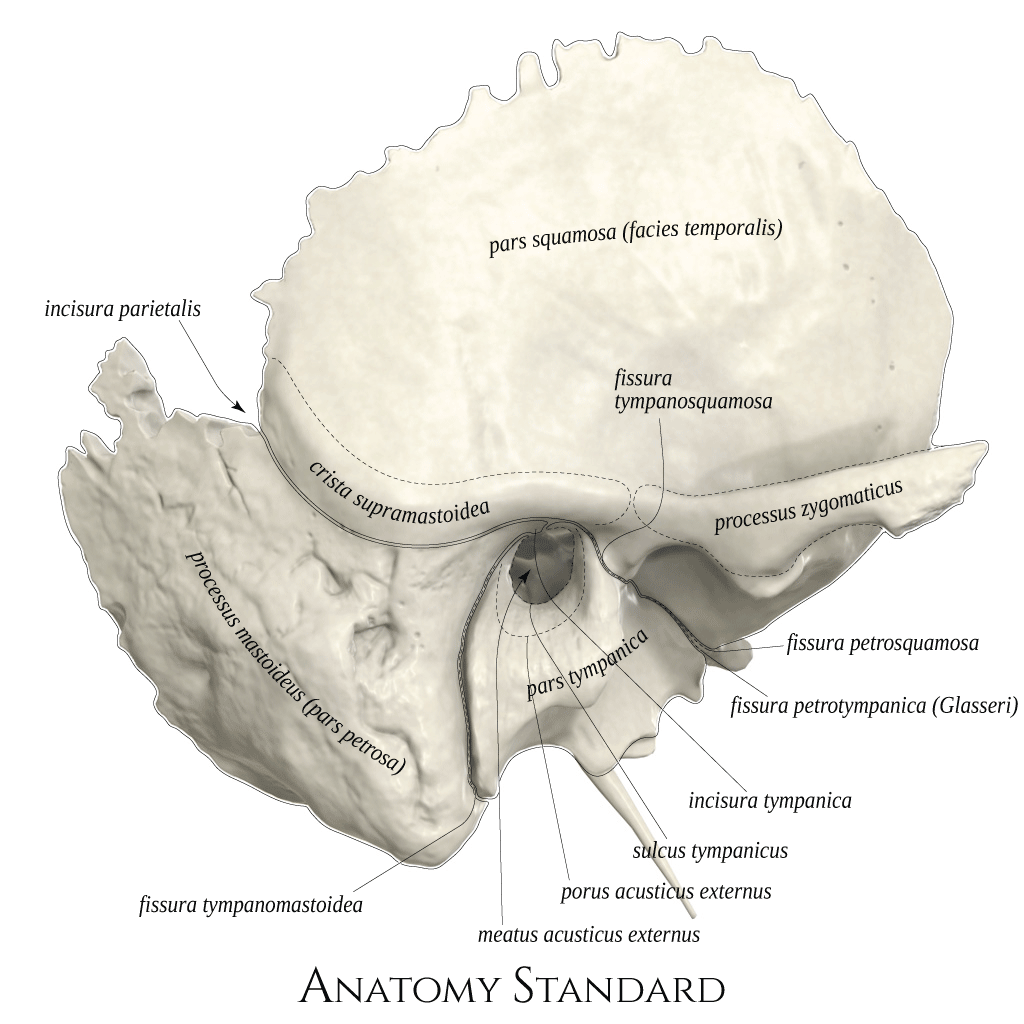
Anatomy Standard Drawing Temporal bone lateral view Latin labels
Case Discussion. Normal CT of the petrous temporal bones. The inner ear structures, ossicles and facial nerves are well demonstrated.
Petrous Portion Of Temporal Bone slidesharedocs
The non-pneumatized petrous apex will show fatty marrow appearing hyperintense on routine T1- and T2-weighted sequences with no expansion of the bone. Confirmation is made by observing the complete loss of signal with fat-saturation sequences. Asymmetric fatty infiltration of the apex may be observed as a conspicuous asymmetric high signal on.
petrous temporal bone
The petrous temporal bone has a pyramidal shape with an apex and a base as well as three surfaces and angles: apex ( petrous apex) directed medially; articulates with the posterior aspect of the greater wing of the sphenoid and basilar occiput. forms internal border of the carotid canal and the posterolateral boundary of the foramen lacerum.
Temporal bone anatomy and labeled diagram GetBodySmart
The squamous part is the anterior superior portion of the temporal bone that forms the lateral part of the middle cranial fossa.It has the appearance of a large flattened plate. Its external surface is smooth and slightly convex. Above the external acoustic meatus, there is a groove on the external surface of the bone for the middle temporal artery.The internal surface of the squamous part is.

Section Through Temporal Bone ClipArt ETC
Attention is called to a little known cranial ossicle, the os supra petrosum (O.S.P.), located within the dura at the tip of the petrous bone. It is an anatomic variant which may be seen quite clearly in the roentgenograms. It is of no apparent clinical significance but should be recognized in the differential diagnosis of intracranial calcifications. Five children with this roentgen finding.